Customize Consent Preferences

We use cookies to help you navigate efficiently and perform certain functions. You will find detailed information about all cookies under each consent category below.

The cookies that are categorized as "Necessary" are stored on your browser as they are essential for enabling the basic functionalities of the site. ... 

Always Active

Necessary cookies are required to enable the basic features of this site, such as providing secure log-in or adjusting your consent preferences. These cookies do not store any personally identifiable data.

No cookies to display.

Functional cookies help perform certain functionalities like sharing the content of the website on social media platforms, collecting feedback, and other third-party features.

No cookies to display.

Analytical cookies are used to understand how visitors interact with the website. These cookies help provide information on metrics such as the number of visitors, bounce rate, traffic source, etc.

No cookies to display.

Performance cookies are used to understand and analyze the key performance indexes of the website which helps in delivering a better user experience for the visitors.

No cookies to display.

Advertisement cookies are used to provide visitors with customized advertisements based on the pages you visited previously and to analyze the effectiveness of the ad campaigns.

No cookies to display.

Pseudoaneurysm after Transradial Coronary Angiography

Juan José Vidal Insua

8 de marzo de 2017

Juan C. Moy Petersen, M.D., and Juan J. Vidal Insua, M.D.

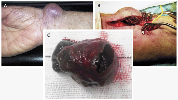
An 81-year-old woman presented with a 1-month history of a large, painless, and pulsatile swelling of the right wrist that had progressively increased in size (Panel A).


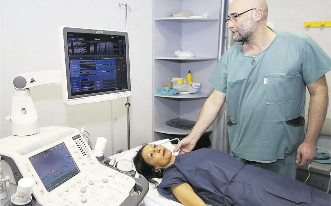
She had a history of hypertension and mitral insufficiency and was taking aspirin, an antihypertensive medication, and a statin. The patient had undergone coronary angiography with a transradial approach 1 month earlier; a 4-French sheath was used, and sheath removal was followed by mechanical clamp compression. A clinical diagnosis of radial pseudoaneurysm was confirmed on ultrasonography. Pseudoaneurysms occur infrequently as a complication of transradial coronary angiography. Predisposing factors include the use of a large catheter sheath, multiple punctures at the site, the use of antiplatelet and antithrombotic agents, inadequate hemostasis or postprocedure compression, and vascular-site infection. The patient underwent surgical excision of the pseudoaneurysm and suture of the radial artery (Panels B and C). She remained well at follow-up visits both 1 week and 2 months after the procedure, with preserved radial arterial pulse and no residual swelling.

Últimas entradas